Exploring the Front Surface of the Eye: Insights and Functions
Written on
Chapter 1: Understanding the Eye's Anatomy
The human eye is often described as a portal to the soul, a captivating organ that enables us to perceive the world in vivid detail. Despite the intricate nature of the visual system, it's the front surface of the eye that is pivotal in our ability to see with clarity. This surface comprises several key structures: the sclera, cornea, conjunctiva, and eyelids, all of which serve protective and focusing roles.
Among the components we will examine are:
- Sclera
- Cornea
- Conjunctiva
- Tear Film
- Lacrimal Apparatus
Let’s delve into the sclera and conjunctiva.
Section 1.1: The Sclera and Conjunctiva
The sclera, commonly recognized as the eye's white part, forms the outer shell that encompasses the eye's inner structures. Composed of collagen and elastic fibers, it is enveloped by a thin membrane known as the conjunctiva (pronounced CON-junk-tie-va).
The conjunctiva is rich in blood vessels and serves crucial functions, including infection prevention and corneal injury healing. When faced with potential threats, the blood vessels within the conjunctiva expand, allowing leukocytes, nutrients, and antibodies to enter the tear film. This tear film nourishes and cleanses the cornea.
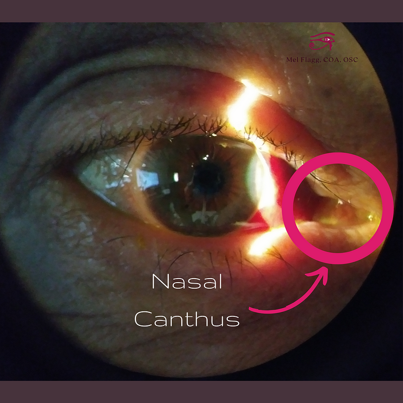
Additionally, the conjunctiva secretes mucus and oils into the tear film, maintaining moisture in the cornea and preventing dryness. The mucus acts as a net to trap microorganisms, which are then directed to the nasal canthus, the area closest to the nose where the upper and lower puncta drain the tear film.

Subconjunctival Hemorrhages
A subconjunctival hemorrhage (SCH) occurs when a blood vessel in the conjunctiva ruptures. Although it may appear alarming, as shown in the image above, these occurrences are generally harmless. The most common triggers include sneezing or coughing, often following illnesses like the flu or allergies. Many individuals remain unaware of their SCH until it is pointed out by someone else.
Typically painless, these hemorrhages can affect anyone but are more frequent among individuals with diabetes, high blood pressure, or those taking blood thinners. While SCHs require no treatment and will resolve in two to three weeks, recurring episodes should prompt a consultation with a healthcare provider to rule out underlying issues.
Section 1.2: The Tear Film
At the heart of protecting the eye's front surface is the tear film, a delicate and dynamic layer composed of water, proteins, lipids, and mucins. This film is essential for maintaining eye health and ensuring visual clarity.
The tear film consists of three layers:
- Lipid Layer: The top layer prevents evaporation and is secreted by the meibomian glands, located at the edge of the eyelids. Occasionally, these glands can become clogged, leading to conditions like chalazion or hordeolum (styes), which are treated with warm compresses and lid scrubs.
- Aqueous Layer: The middle layer, produced by the lacrimal glands beneath the eyebrow, hydrates the cornea and provides essential nutrients.
- Mucous Layer: This innermost layer is secreted by goblet cells in the conjunctiva, facilitating oxygen transport and smoothing the corneal surface for optimal refraction.
The Lacrimal System
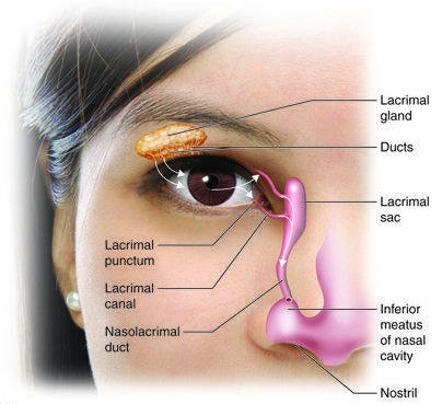
Next, we explore the lacrimal system, which drains the tear film. Tears flow through the punctum (small drains in the eyelids), into the lacrimal sac, and down the nasal lacrimal duct, explaining why crying often leads to a runny nose.
In rare cases, obstructions in the lacrimal duct can cause infections, known as dacryocystitis, requiring flushing or probing for resolution.

Eyelashes
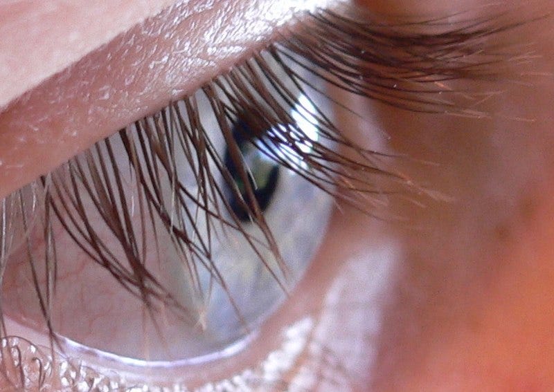
Eyelashes serve a vital role beyond aesthetics; they act as a barrier against dust and debris. Sensitive to touch, they trigger reflexive eye closure when approached by objects.
Eyelashes undergo a natural cycle, shedding one to five per day, and can be affected by conditions like blepharitis, which causes itching and inflammation. Treatment often involves lid scrubs and, in some cases, medicated drops.

Conclusion
The eye's front surface, with its intricate structures and systems, plays a critical role in vision and overall ocular health. Understanding these components can help us appreciate the delicate balance required for maintaining clear sight.
The first video, "PUSHA T - OPEN YOUR EYES," explores the importance of awareness and perception, aligning with our insights into the eye's anatomy.
The second video, "How to Get Rid of Eye Floaters," provides practical advice on managing common eye concerns, complementing our discussion on ocular health.